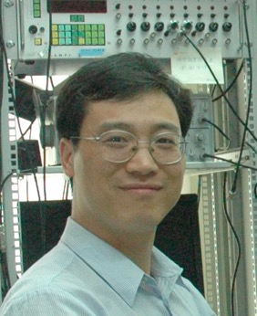

Yousheng Shu, Ph.D.
EDUCATION
B. S. 1990-1994 Biology
Department of Biology, Hunan Normal University
Ph.D. 1994-1999 Neurobiology and Neuropharmacology
Shanghai Brain Research Institute, Chinese Academy of Sciences
PROFESSIONAL TRAINING
1999-2004 Postdoctoral associate
2004-2006 Associate research scientist
Department of Neurobiology (and Department of Anesthesiology), Yale University
APPOINTMENTS
2006-2013 Principal Investigator, Head of the Lab of Neural Network Function Institute of Neuroscience, Chinese Academy of Sciences
2013-present Full Professor, Dean
School of Brain and Cognitive Sciences
State Key Laboratory of Cognitive Neuroscience and Learning
Beijing Normal University
AWARDS AND HONORS
1. The “Sanofi Aventis-SIBS Young Faculty Award” (2009)
2. The “Outstanding Young Scientist Award” of CAS Shanghai Branch (2010)
3. The “Young Scientist Award” of Chinese Academy of Sciences (2011)
4. The “Outstanding Supervisor of Graduate Students” Award (2012)
ACADEMIC AND PROFESSIONAL SERVICES
Referee for the following journals:
Acta Pharmacologica Sinica; Acta Physiologica Sinica; Biochemical and Biophysical Research Communications; Biophysical Journal; Cerebral Cortex; Chinese Journal of Cell Biology; European Journal of Neuroscience; Frontiers in Computational Neuroscience; Frontiers in Neural Circuits; Journal of Neuroscience; Journal of Neurophysiology; Journal of Physiology; Nature Reviews Neuroscience; Neuroscience Bulletin; PLoS Biology; PLoS ONE; PNAS; Science in China
MEMBERSHIPS
1. Member of Society for Neuroscience USA (1999-present)
2. Member of Chinese Society for Neuroscience (1997-present)
SUMMARY OF CURRENT RESEARCH
Research direction of the lab: Mechanisms of Signal Processing in Cortical Neurons and Microcircuits. The long-term goal of our research is to understand how individual neurons and their interconnected local networks contribute to functioning of the cerebral cortex. At the subcellular and cellular level, we seek to reveal the mechanisms of synaptic integration, action potential (AP) initiation (neuronal excitability) and neural coding; while at the circuit level, we aim to reveal the mechanisms by which the cortical circuit generates its variety of activity patterns. The objective of our recent studies was to characterize the biophysical properties of axonal ion channels in excitatory pyramidal cells (PCs) and inhibitory interneurons, and to investigate how these properties could be regulated by neuromodulators. Using human cortical tissue discarded during surgical removal of brain tumors and epileptic seizure foci, we search for special cell types in the human cortex that may have distinct morphological or electrophysiological properties, as well as unique microcircuits in the human cortex. In addition, we study the cellular and circuit mechanisms for the development of brain disorders, particularly those with imbalanced excitation and inhibition such as anxiety and epilepsy. The following statements conclude the research we have done since the lab was set up in China in 2006.
1. Mechanisms for the generation of digital and analog signals in cortical neurons
Cortical neurons communicate in both digital and analog modes (Shu et al., 2006, 2007a). The digital signal is the all-or-none APs initiated first at the axon initial segment (AIS). The analog mode of communication reflects the modulation of synaptic strength by presynaptic membrane potential (Vm). The average amplitude of AP-triggered postsynaptic responses increases with presynaptic depolarization. We investigated the underlying mechanism for the two modes of communication.
For the generation of digital signals, we performed patch-clamp recordings from cortical axons (i.e. tight-seal axonal bleb recording, developed by Shu et al., 2006, 2007) in brain slices, together with immunostaining experiments, and found that different subtypes of voltage-gated Na+ channels play distinct roles in mediating AP initiation and propagation. In excitatory pyramidal cells (PCs), subtype Nav1.6 accumulated at the distal end of AIS mediates AP initiation, whereas Nav1.2 concentrated at the proximal AIS promotes AP backpropagation to the soma and dendrites (Hu et al., 2009). In inhibitory interneurons, we found the expression of proximal Nav1.1 and distal Nav1.6 at the AIS of parvalbumin-containing neurons. In addition to these channel subtypes, we also observed the expression of Nav1.2 at the AIS of somatostatin-positive interneurons. The distinct channel subtype compositions contribute to the difference in AP thresholds in the two cell types. The presence of Nav1.2 in axons of cortical inhibitory interneurons was surprising because it has been long believed that this channel subtype only exists in PCs. However, our result explains well clinical observations that both gain- and loss-of-function mutations of Nav1.2 increase the susceptibility of epilepsy. Indeed, blocking Nav1.2 pharmacologically promotes the generation of epileptiform activity in Mg2+-free ACSF (Li et al., 2014).
For analog signaling, our axon recording revealed a selective expression of a rapidly activating but slowly inactivating K+ current. Pharmacological experiments demonstrated that this current is mediated by Kv1 channels. Subthreshold depolarization of the axon, but not the soma, causes rapid activation of this current followed by slow inactivation with a time constant of 6-7 sec. This time constant is similar to that of depolarization-induced changes in AP width and enhancement of synaptic transmission, suggesting a role of Kv1 channels in mediating the Vm-dependent modulation of synaptic transmission (Shu et al., 2007b). Therefore, our results provide a new mechanism for analog-mode communication between cortical neurons.
2. Regulation of axonal ion channel and AP waveform by neuromodulators
We investigated whether axonal ion channels are subjected to regulation by neuromodulators such as dopamine and serotonin. We found that K+ currents recorded at the PC axons could be reduced and enhanced by D1 and D2 receptor agonists, respectively. Consistently, the width of axonal AP increased upon the activation of D1 receptors. We further showed that the modulation also occurred in axons isolated from the soma, indicating the expression of dopamine receptors in cortical axons (Yang et al., 2013). This study provides a new mechanism for dopaminergic modulation of neuronal signaling and synaptic transmission. In a different study, we observed failures of AP backpropagation from the axon to the soma in PCs upon the activation of 5-HT1A receptors. Further experiments revealed that axonal Na+ currents mediated by Nav1.2 but not Nav1.6 are inhibited by 5-HT1A receptor activation (Yin et al., 2015). This selective modulation controls AP backpropagation to the somatodendritic compartments, possibly modulating synaptic plasticity that depends on the timing of backpropagating APs.
3. Regulation of microcircuit function by analog-mode communication
A dynamic balance of excitation and inhibition is crucial for network stability and cortical processing, it remains unclear how the cortex achieves this balance at various cortical states. We reasoned that analog-mode communication between excitatory neurons may also occur at synapses from excitatory to inhibitory neurons, and the amount of recurrent inhibition might be subject to change in a Vm state-dependent manner. Using paired recording from PCs and inhibitory interneurons, we indeed revealed a state-dependent modulation of recurrent inhibition between pyramidal cells (PCs) mediated by two distinct interneuron subtypes, low-threshold spiking (LTS) and fast-spiking (FS) cells. PC depolarization (increased excitability) enhanced the amplitudes of AP-triggered EPSPs in interneurons, and thus increased their probability of spiking or number of APs, which would subsequently provide more inhibition to neighboring PCs (Zhu et al., 2011). These results show a critical strategy of the cortex to dynamically control the excitation-inhibition balance in the network. The results also demonstrate that the analog signaling has profound impact on the operation of cortical circuits.
4. Changes in intrinsic property and synaptic transmission in epileptic cortical tissue
Normally, neurotransmitter release is tightly coupled or synchronized with presynaptic AP generation. However, prolonged asynchronous release (AR) of transmitter for hundreds of milliseconds following presynaptic AP burst has been observed at some excitatory and inhibitory synapses, particularly after high-frequency firing of presynaptic neurons. At GABAergic synapses, AR may provide long-lasting inhibition and reduction of discharge probability and precision in postsynaptic neurons, leading to desynchronization of network activities. We found that AR occurs at output synapses of FS neurons, including FS autapses, FS-FS and FS-PC synapses in the human neocortex. Surprisingly, in comparison with that in non-epileptic tissues, the AR strength is stronger in epileptic tissues, possibly resulting from an upregulation of Na+ channel expression in FS neurons (Jiang et al., 2012). We further demonstrated the existence of AR in FS neurons in rat cortex, and the AR strength in juvenile animals is stronger than that in adults. The strength transition may occur during the critical period of prefrontal cortical functions (Jiang et al., 2012, 2015). These results, to our knowledge, provide the first piece of evidence showing the occurrence of GABAergic AR in human tissue, and importantly reveal an increase in AR in the epileptic neocortex, suggesting a role of AR from FS neurons in regulating the synchrony of network activities and thus shaping epileptiform activities.
5. A distinct subpopulation of inhibitory interneuron in human and monkey cortex
Neuronal types such as PCs and non-pyramidal interneurons in the neocortex are highly conservative during evolution. It remains unclear whether human neurons have physiologically distinct features. We performed whole-cell recording from acute human cortical slices and found a novel type of persistent-activity neuron (PAN). The persistent activity in these neurons is intrinsic (independent of synaptic activity) and can be turned on by weak (single AP) but turned off by strong stimulation (AP burst). Further investigation showed that it is attributable to a depolarizing plateau potential induced by a persistent Na+ current. Single-cell RT-PCR results indicate that these neurons are inhibitory interneurons mainly containing somatostatin, calretinin or cholecystokinin. Among cortical interneurons, the estimated percentage of these neurons was approximately 9%. PANs were also observed in monkey but not rat cortical tissues, suggesting they could be unique to primates (Wang et al., 2015a and b). The characteristic persistent activity in these inhibitory interneurons may contribute to the regulation of PC activity and participate in high-order brain function.
BOOK CHAPTERS
1. McCormick DA, Shu Y, and Hasenstaub A (2003) Balanced recurrent excitation and inhibition in local cortical networks. Book chapter in Excitatory-inhibitory balance: synapses, circuits, and systems plasticity. Edited by T. Hensch. Kluver Academic Press.
2. Shu Y (2008) Recurrent Cortical Network Activity and Modulation of Synaptic Transmission. Book chapter in Seizure Prediction in Epilepsy. Edited by B. Schelter, J. Timmer and A. Schulze-Bonhage. WILEY-VCH Verlag GmbH & Co. KGaA, Weinheim.
3. Hu W and Shu Y (2012) Axonal Sodium Channels and Their Role in Action Potential Initiation and Propagation. Book chapter in Progress in Physiological Sciences. (Chinese)
PUBLICATIONS
1. Wang T, Yin L, Zou X, Shu Y, Rasch M, Wu S. (2016) A Phenomenological Synapse Model for Asynchronous Neurotransmitter Release. Front. Comput. Neurosci. Doi: 10.3389/fncom.2015.00153
2. Yin L, Rasch M, He Q, Wu S, Dou F, Shu Y. (2015) Selective Modulation of Axonal Sodium Channel Subtypes by 5-HT1A Receptor in Cortical Pyramidal Neuron. Cerebral Cortex. Doi: 10.1093/cercor/bhv245
3. Wang B, Shu Y. (2015) Physiologically Distinct Neuronal Type in Primate Cortex. Oncotarget. Doi: 10.18632/oncotarget.4796
4. Wang B, Yin L, Zou X, Ye M, Liu Y, He T, Deng S, Jiang Y, Zheng R, Wang Y, Yang M, Lu H, Wu S and Shu Y. (2015) A Subtype of Inhibitory Interneuron with Intrinsic Persistent Activity in Human and Monkey Neocortex. Cell Reports, Doi.org/10.1016/j.celrep.
5. Jiang M, Yang M, Yin L, Zhang X, and Shu Y. (2015) Developmental reduction of asynchronous GABA release from neocortical fast-spiking neurons, Cerebral Cortex. 25(1):258-70.